PRÉSENTATION DES RÉSULTATS DE LA RECHERCHE
Le protocole FLUPROSTIC
Rationnel
La FCH est un MRP plus performant que le FDG, traceur de référence actuellement le plus utilisé en TEP clinique oncologique, en particulier pour le bilan d’extension initial ou de la récidive du cancer de la prostate. La FCH est également plus performante que le FNa pour la détection des métastases osseuses du cancer de la prostate. Cependant, la FCH est aussi plus chère à l’achat pour l’hôpital que le FDG ou le FNa, alors que son remboursement par l’assurance maladie est identique. La place respective de la FCH et du FNa dans la stratégie thérapeutique du cancer de la prostate reste donc à définir.
C’est pour tenter de répondre à cette question qu’a été proposée l’étude FLUPROSTIC, « Place de la TEP/TDM au fluor-18 et de l’IRM de diffusion « corps entier » dans la détection des premières métastases osseuses du cancer de la prostate », étude française multicentrique prospective ouverte aux inclusions entre décembre 2011 et août 2014. Ce protocole de recherche a été réalisé dans le cadre du programme « Soutien au Thérapies Innovantes et Coûteuses » (STIC), désormais appelé Programme de Recherche Médico-Economique (PRME), sur des fonds accordés par le Ministère des Solidarité et de la Santé (STIC 2009 – P090105) et promu par l’Assistance Publique – Hôpitaux de Paris (NCT01501630). L’objectif médical principal était de comparer prospectivement les performances diagnostiques de la TEP/TDM au FNa, la TEP/TDM à la FCH et de l’IRM du « corps entier » avec séquences de diffusion dans la détection des métastases osseuses du cancer de la prostate. Les objectifs médico-économiques étaient une comparaison de leurs impacts respectifs sur la décision thérapeutique dans des conditions de « vie réelle » et une comparaison coût-efficacité des différentes stratégies d’imagerie.
Population
Les patients avec un diagnostic de cancer de la prostate prouvé histologiquement, suspects d’êtres métastatiques au niveau de l’os, étaient éligibles. Ils pouvaient être en situation de bilan d’extension initial, de récidive biologique, ou de résistance à la castration. Les critères de suspicion de métastases osseuses étaient un score histopronostic de Gleason supérieur à 8, un stade initial T3b ou supérieur, une durée de survie sans récidive biologique inférieure à 3 ans après traitement initial curatif, un temps de doublement de la concentration sérique du PSA inférieur à 3 mois. Après obtention du consentement éclairé signé, les patients étaient programmés pour passer les trois modalités d’imagerie étudiées dans un ordre randomisé, en plus du bilan standard du cancer de la prostate recommandé selon la situation clinique, dans un délai maximal de 1 mois sans modification thérapeutique dans cet intervalle.
Cinq centres ont participé au recrutement des patients et l’imagerie a été réalisée dans deux centres spécialisés en imagerie oncologique (l’hôpital Tenon à Paris et l’institut de cancérologie de l’ouest René Gauducheau à St-Herblain).
Après avoir complété le bilan d’imagerie, chaque patient a été traité et suivi par son clinicien référent en fonction des recommandations en vigueur de l’Association Française d’Urologie.
La durée minimale du suivi devait être de 6 mois pour établir le standard de vérité pour les performances de l’imagerie, et de 12 mois pour l’analyse médico-économique afin d’obtenir les coûts de traitements réels.
Standard de vérité pour les métastases osseuses
La présence de lésions osseuses a été quottée selon plusieurs régions et résumée dans une approche par-patient (présence ou absence d’atteinte osseuse). La réalité de l’atteinte osseuse a été déterminée par un panel de spécialistes (un urologue, un radiothérapeute, un radiologue et un médecin nucléaire) n’ayant pas participé à l’essai FLUPROSTIC, tous avec au moins 10 ans d’expérience dans le cancer de la prostate. Le standard de vérité a été réalisé pour chaque patient après évaluation de l’ensemble des éléments cliniques, biologiques, histologiques et d’imagerie disponibles dans le suivi clinique. Les vrais positifs de l’imagerie étaient définis en premier lieu sur la concordance des 3 modalités du protocole, et en cas de discordance, sur les éléments clinico-biologiques de suivi, en particulier la réponse du PSA à des traitements ciblés à visée curative (baisse du PSA d’au moins 50% comparativement à la valeur de base), en excluant les traitements de déprivation androgénique. Les vrais négatifs de l’imagerie étaient définis par une absence d’évolutivité osseuse à 6 mois sur l’ensemble des données de suivi disponible.
Materials and methods
Population
Patients with histologically proven PCa and without a known history of metastases were eligiblefor the French multicentre study “FLUPROSTIC” (NCT01501630) aiming to compare the utility of NaF-PET/CT, FCH-PET/CT and DW-MRI for detecting unknown PCa metastasis. Inclusion was offered to any PCa patient suspicious for distant metastasis referred by their clinician in any of the following settings: initial staging, biochemical recurrence (BCR) without ongoing androgen-deprivation therapy (ADT), biochemical recurrence during ADT, corresponding to castration-resistant prostate cancer (CRPC). Patients were considered at risk of developing metastases if they were presenting any of the following situations: Gleason score 8–10, time to BCR less than 3 years, stage T3b or higher, or a PSA doubling time less than 3 months (for BCR and CRPC) (18,104,111). Patients presenting suspicious bone pain were also considered.
BCR was diagnosed after surgery, radiotherapy or alternative local treatment options with curative intent according to current recommendation for BCR and CRPC patients, respectively (16). In addition to the standard PCa workup, patients underwent those three WBIs within less than a month without any therapeutic intervention during this period of time. Exclusion criteria were other progressive neoplasias, therapy change during the imaging workup, WBI workup not completed within 1 month, and contra-indications to any of the imaging modalities.
Five centres recruited patients. WBI was performed in two centres that specialised in cancer imaging (Hôpital Tenon, Paris and ICO René Gauducheau, St-Herblain).
Patient follow‑up and adequacy of management
The median duration of follow-up was 28 months (range 6–70). Five patients were lost to follow-up and excluded from this analysis; WBI performed at staging (n = 1), BCR (n = 3) or CRPC (n = 1) had an impact on the management in 2 of them. Eighty of the 101 patients were evaluable for bone metastases (21 staging, 48 BCR and 11 CRPC) according to a composite standard of truth based on the evolution of the lesion on imaging during follow-up, evolution of PSA in response to targeted prostate cancer therapy, excluding ADT and histological findings, if available. At least one bone metastasis was found in 14/80 patients at the time of WBI: 5/21 staging patients; 7/48 BCR patients and 2/11 CRPC patients.
At the time of database lock, 26/26 patients referred for staging were PCa recurrence–free; 46/56 BRC patients had no scalable disease whereas 10/56 presented with continuous PCa evolution; 9/14 CRPC patients had no scalable disease, whereas 5/14 had progressive disease.
The global adequacy rate of management decisions was 98% (95% CI 95–100%). Of the 27 assessable patients in whom WBI had a clinical impact, the changes were considered as adequate in 26/27 = 96% (95% CI 89–100%) of patients but inadequate in one single case: in a high-risk (according to d’Amico) patient initially treated by prostatectomy, who was planned for ADT due to his first BCR event (serum PSA 0.9 ng/mL), suspicious pelvic lymph nodes were detected on FCH-PET/CT and dissected. Pathology results were negative and serum PSA level continued to rise to 4.57 ng/mL 6 months after surgery. ADT was finally introduced whichled to a decrease in PSA serum level (0.51 ng/mL 3 years after FLUPROSTIC workup).
The management of the 68 assessable patients in whom WBI had no impact was considered adequate in all patients but one: in a high-risk patient initially treated by HIFU presenting with his first BCR event (serum PSA 16.5 ng/mL), resuming HIFU was planned and performed because an isolated prostate lesion was detected on imaging. HIFU was complicated by infection and prostate necrosis which required hospitalisation and was followed by a rapid increase in serum PSA level to 92 ng/mL. 8 months after the WBI workup, a bone metastasis was detected on MRI of the pelvis.
Discussion
To the best of our knowledge, this report describes the first direct comparison of the impact of NaF-PET/CT, FCH PET/CT and DW-MRI findings on the management of PCa patients referred for suspected metastasis at three different phases of evolution. WBI with those three modalities prompted management changes in 29% of patients, which were considered as adequate in all patients but two. If only one single WBI examination would have been performed in our 101 patients, the impact rate would have been 28% with FCH-PET/CT, 6% with NaF-PET/CT and 9% with DW-MRI. This result confirms, on a rational basis, the observation that introducing FCH-PET/CT for routine PCa WBI results in a decrease in demand for nuclear imaging of the skeleton (NaFPET/CT and bone scintigraphy) (47).
According to the evolution status of PCa, the impact at staging was of 2/27 patients for each of the three WBI, which showed bone metastases in the same two patients. Hillner et al. retrospectively evaluated the impact rate of NaF-PET (without CT fusion) to be 12.2% at staging in 1024 PCa patients (45). In the study of Kjölhede et al. on 90 patients, the impact rate of combined NaF and FCH PET/CTs on first-line treatment plan was 20% (56). The impact of FCH and DWI-MRI performed for staging PCa was never evaluated. However, in our study, changes in management at staging were most frequently decided independent of WBI (Table 2). Our main finding was that WBI had its highest impact rate in BCR, 41% overall and 39% for FCH-PET/CT, significantly superior to NaF-PET/CT or DW-MRI (p < 0.0001). This impactrate of 39% is within the confidence limits of those reported by Soyka et al. (48% on 156 patients) (98) and Gillebert et al. (56% on 179 patients) (40). Using 11C-choline-PET/CT, Ceci et al. reported an impact rate of 46.7% on 150 patients (12). In 15 CRPC patients, we found no statistically significant difference between the impact of the three WBI, only a trend for a greater rate of FCH-PET/CT (3/15 patients vs. 1/15 for DW-MRI and 0/15 for NaF-PET/CT). To the best of our knowledge, the impact of WBI in CRPC has never been reported.
An interesting finding was that the planned management was changed at the post-WBI multidisciplinary meeting independent of the results of WBI in 23% of patients. We assumed thatthese changes were motivated by rethinking the risk of metastatic evolution of PCa duringthat time interval. In 8/23 of these cases the decision was based on serum PSAchanges.
Limitations
A limitation, common to studies comparing impact on management is that the decisions are made on the basis of on-site reading. Therefore, carry-over of information between the reports of the WBI may exist: the latter examination can have an advantage. However, we found no significant difference in the order of performing the three WBI. Furthermore, FCH-PET/CT, which had the highest impact overall and in the BCR group, was performed first in a large proportion of patients (Table 1), which rules out systematic carry-over information from the two other WBI.
Another limitation is the relatively small number of patients and the heterogeneity of their PCa status (staging, BCR, CPRC). However, we did not aim to compare the results between the three groups of patients as they did not present the same risk for metastatic disease, but to cover different settings of PCa, reflecting the prescription of the clinicians. Furthermore, the impact that rate we found for NaF or FCH-PET/CT was similar to those previously reported at staging or for BCR in larger non-comparative series.
There is a difference between WBIs in the actual field of view: a real whole-body for FNaPET/CT (similarly to bone scintigraphy) vs. from vertex to mid-thigh for FCH-PET/CT and DW-MRI. However, no abnormality suggestive of “peripheral” bone metastasis in the limbs was discovered on NaF-PET/CT, confirming that bone metastases of PCa are predominant in the axial skeleton (62) and validating WBI from vertex to mid-thigh.
According to “local habits”, imagers and clinicians more familiar with one WBI modality might have been more prone to “believe it” and implement changes guided by this modality. Indeed, in both imaging centres, clinical trials evaluating FCH-PET/CT were performed for several years (40,47,60), whereas NaF and DW-MRI were introduced on the year before the start ofFLUPROSTIC, possibly contributing to their lower impact compared to FCH-PET/CT. Modern WBI is evolving rapidly. For DW-MRI, a b value of 600 was used, which is below the currently recommended values of at least 800, probably limiting its impact. In this study, MRI was performed without contrast enhancement, which does not contribute toimproved performances of DW-MRI, which was the MRI sequence that we aimed to evaluate.Thus, we chose to not perform contrast-enhancement to avoid a potential adverse reaction of gadolinium contrast media.
Prostate-specific membrane antigen (PSMA) radioligands (26) were not available when FLUPROSTIC was started. PET/CT appears to yield better diagnostic performance with PSMA radiotracers than with FCH (2), with a significant impact on patients’ management (35,36,73).However, PSMA ligands are not currently approved and are not as widely available as FCH inthe EU.
Conclusions
Within the limits of a relatively small series of PCa patients suspicious for distant metastasis where imaging was performed early in the disease transition, we found that WBI led to management changes in approximately 1/3 of cases. For BCR, FCH-PET/CT had the greatest impact rate and may therefore be recommended as first-line WBI. For staging PCa, FCH should be considered instead of NaF-PET/CT, as is currently recommended in clinical guidelines, since it provides additional information on extra-osseous extension of PCa, but this is also true for WB-MRI, which has the advantage of avoiding radiation exposure at this early stage of evolution. Prescribing WBI at a multidisciplinary meeting led to management changes at the post-WBI meeting independent of WBI results in approximately 1/4 of the patients as a consequence of rethinking and of the evolution of PCa during that time interval. These results warrant further investigation. In particular, exploring management changes induced by WBI according to PSA serum levels, which are correlated with imaging performances, will be of interest.
Résumé des résultats
Les performances de la TEP/TDM au FNa, de la TEP/TDM à la FCH et de l’IRM « corpsentier » avec séquences de diffusion dans la détection des métastases osseuses du cancer de la prostatesont connues et publiées. Dans l’article précédent, nous avons comparé leur impact sur la décision thérapeutique, impact particulièrement marqué en situation de première récidive biologique. Dans ce second article nous avons effectué une analyse coût-utilité comparant les trois modalités d’imageries du protocole FLUPROSTIC dans la détection des métastases osseuse chez les patients présentant une première récidive biologique de cancer de la prostate, en nous plaçant de la perspective de l’assurance maladie.
Nous avons construit un modèle combinant un arbre décisionnel intégrant les performances diagnostiques des techniques d’imageries évaluées dans le protocole FLUPROSTIC, sommées dans une approche « par-patient », et un modèle de Markov basé sur l’histoire naturelle du cancer de la prostate afin d’extrapoler les résultats sur un horizon temporel « à vie ». Le modèle de Markov contenait cinq états de santé: récidive biologique avec et sans métastase osseuse, résistance à la castration avec et sans métastase osseuse et mort. La durée des cycles du modèle était d’un an. Les coûts des traitements étaient ceux des patients inclus dans FLUPROSTIC, extraient de l’étude nationale des coûts pour les coûts hospitaliers et des données de l’assurance maladie pour les coûts des traitements en externe. Les probabilités de transition et les utilités utilisées dans le modèle de Markov ont été extraites de la littérature.
Nous n’avons pas retrouvé de différence statistiquement significative entre les trois modalités d’imagerie dans leur performance à détecter des métastases osseuses chez les patients en première récidive biologique de cancer de la prostate, mais la TEP/TDM à la FCH était la plus exacte. La TEP/TDM à la FCH était la modalité d’imagerie la plus coût-utile avec un RDCR de 993€ par QALY gagné si l’interprétation de l’imagerie était faite par des médecins spécialistes locaux, non masqués des données cliniques, et de 5055€ par QALY gagné sil’interprétation était faite par des experts, masqués des données cliniques. Ainsi, la TEP/TDMà la FCH avait 100% de chance d’être la stratégie d’imagerie la plus coût-utile pour un seuil de volonté à payer de 3000€ ou 9000€ par QALY gagné, si l’interprétation de l’imagerie était faite par des médecins spécialistes locaux ou des experts respectivement. De la perspective de l’assurance maladie, la TEP/TDM à la FCH devrait donc être recommandée pour le bilan d’extension des patients en première récidive biologique de cancer de la prostate.
La perspective de l’hôpital n’a pas été développée dans cet article, mais les données des coûts ont été collectées pour l’activité de l’hôpital Tenon en 2018 et 2019. Actuellement en France, les examens TEP/TDM sont remboursés de la même manière par l’assurance maladie, quel que soit le MRP, correspondant à 89.54€ d’acte d’interprétation de l’imagerie TEP (code CCAM ZZQL016) plus un forfait technique reversé au centre d’imagerie de 1000€ pour les 1000 premiers examens de l’année et de 550€ pour les examens suivants (NOR: AFSU1704105S; JORF n°0037 du 12 février 2017 – texte n° 17). Considérons la machine TEP/TDM la plus récente du service de médecine nucléaire de l’hôpital Tenon. Nous ne prendrons pas en compte les coûts de structure, identiques pour toutes les imageries. Nous considèrerons aussi que les examens TEP/TDM ont été réalisés régulièrement et de manière homogène au cours de l’année.
En 2018, cette machine TEP/TDM a réalisé en routine clinique 3375 examens TEP (sans compter les explorations TEP cérébrales), dont 68 avec du FNa et 389 avec de la FCH pour exploration de cancer de la prostate (toutes indications confondues). Les coûts de production des examens TEP/TDM était de 303€ pour le FNa et 882€ pour la FCH, pour des coûts du MRP de 164€ et 743€ par examen respectivement (Table additionnelle 4 de l’article). Ainsi, en 2018, les examens TEP/TDM au FNa et à la FCH ont coûté à l’hôpital Tenon 20 604€ et 343 098€ respectivement. Si nous supposons qu’1/3 de la totalité de chaque examen a été remboursé avec le forfait technique de 1000€ et les 2/3 restant avec celui de 550€, les examens TEP/TDM au FNa et à la FCH ont rapporté à l’hôpital Tenon 53 720€ et 307 310€ respectivement, soit un gain de 33 116€ pour le FNa et une perte de 35 788€ pour la FCH.
En 2019, cette même machine a réalisé en routine clinique 3308 examens TEP/TDM, dont 67 avec du FNa et 336 avec de la FCH pour exploration de cancer de la prostate (toutes indications confondues). En 2019, le coût de la FCH a été renégocié par l’hôpital à 600€ par examen, permettant une baisse du coût de production de cet examen à 739€ par examen. Le coût du FNa n’a pas varié. Par conséquent, en 2019, les examens TEP/TDM au FNa et à la FCH ont coûté à l’hôpital Tenon 20 301€ et 248 304€ respectivement. Ces mêmes examens ont rapporté 52 930€ et 265 400€ respectivement, et ont donc rapporté à l’hôpital Tenon 32 629€ et 17 136€ respectivement.
Ainsi, une baisse de 20% du coût de la FCH a rendu la réalisation des TEP/TDM à la FCH rentable pour notre hôpital, renforçant l’intérêt de son utilisation dans le cancer de la prostate.
Nous ne développerons pas l’IRM pour ces résultats complémentaires car elle n’est pas la modalité d’imagerie étudiée dans cette Thèse.
Background
Prostate cancer (PCa) is the second most prevalent cancer in men worldwide, accounting for approximately 15% of all diagnosed cancers (29), presenting an annual incidence of 31.1 per 100,000 cases and being the 3 rd cause of death by cancer in men behind lung and colorectal cancers (29). Bone is the most frequent metastatic site of PCa and is the only metastatic site in approximately 62% of cases (54). The prevalence of bone metastasis in PCa is 3% at diagnosis (for all stages), and its estimated cumulative incidence is 16.6% 5 years after PCa diagnosis (78). The discovery of bone metastases marks a turning point in the history of the disease, especially in terms of treatment strategy and prognosis, and patients with bone metastases have a worse prognosis (78,92,100).
F-sodium fluoride positron emission tomography/computed tomography (PET/CT) (NaF), F fluorocholine PET/CT (FCH) and diffusion-weighted whole-body magnetic resonance imaging (DW MRI) are three novel imaging techniques that are effective for the detection of PCa bone metastases and their results may impact the management of patients (34). Their diagnostic performances in detecting bone metastases in PCa patients with first biochemical recurrence (BCR) have already been published (40,60,109). However, to the best of our knowledge, their cost-effectiveness in this indication has never been compared. The objective of this study was to provide evidence for policy making at the national level by comparing the cost-effectiveness of the detection of bone metastasis in PCa patients with first BCR by means of these three imaging modalities.
Methods
The French multicentre study “FLUPROSTIC” (NCT01501630) was a prospective integrated clinical and economic national multicentre study comparing NaF, FCH and DW-MRI in detecting bone metastasis in PCa patients with first BCR. The medical objective was to compare the diagnostic performance and the impact on patient management of these three imaging modalities in this indication. It was completed by a cost effectiveness analysis using real treatment costs of trial patients. The FLUPROSTIC trial was conducted in accordance with the Declaration of Helsinki and approved by a national review board (IDRCB 2011-A01041–40). All patients provided written informed consent. The study design, inclusion/exclusion criteria, follow-up and standard of truth (SOT) for bone metastasis that were applied in this study are summarized in Additional file 1. The imaging protocols and imaging interpretation are detailed below. For the purpose of economic evaluation, we extrapolated the results of the trial using astate-transition model.
|
Table des matières
INTRODUCTION
PARTIE 1 : ETUDES MÉDICO-ÉCONOMIQUES RAPPORTANT L’INTÉRÊT DE L’UTILISATION DE LA TEP/TDM EN ONCOLOGIE
1.1- Cancer du poumon
1.1.1- Caractérisation de nodule pulmonaire
1.1.2- Bilan d’extension initial du cancer du poumon
1.2- Cancer du sein
1.2.1- Bilan d’extension initial du cancer du sein
1.2.2- Autres indications dans le cancer du sein
1.3- Cancer de la prostate
1.3.1- Dépistage du cancer de la prostate
1.3.2- Bilan d’extension initial du cancer de la prostate
1.3.3- Bilan d’extension de la récidive du cancer de la prostate
1.4- Cancer colorectal
1.5- Autres cancers
PARTIE 2 : PRÉSENTATION DES RÉSULTATS DE LA RECHERCHE
2.1- Le protocole FLUPROSTIC
2.1.1- Rationnel
2.1.2- Population
2.1.3- Standard de vérité pour les métastases osseuses
2.2- Impact de la TEP/TDM au
F-fluoride de sodium, de la TEP/TDM à la F-fluorocholine et de l’IRM corps entier avec séquences de diffusion dans le management des patients ayant un cancer de la prostate suspect d’être métastatique : une étude prospective multicentrique
Résumé des résultats
Introduction
Materials and methods
Results
Discussion
Conclusions
Table 1: Patients characteristics
Table 2: Management plans advised by the multidisciplinary panel before and after imaging workup, and impact of the results of whole-body imaging
2.3- Comparaison de la TEP/TDM au
F-fluoride de sodium, de la TEP/TDM à la F-fluorocholine et de l’IRM corps entier avec séquences de diffusion dans la détection des métastases osseuses chez les patients en récidive de cancer de la prostate : une étude coût-efficacité française
Résumé des résultats
Background
Methods
Results
Discussion
Conclusion
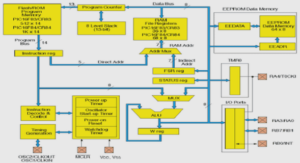
Figure 1 title: Decision tree combined with the Markov model used for evaluating costs and health related outcomes
Figure 2 title: Incremental cost in Euros and effect of imaging strategies on the cost-effectiveness plane
Figure 3 title: Tornado plot presenting uncertainties in costs in Euros within plausible ranges of the 95%confidence intervals
Figure 4 title: Scatter plot showing the uncertainty of the incremental cost-effectiveness ratio in Euros for each imaging modality
Figure 5 title: Cost-effectiveness acceptability curves in Euros showing the results of probabilistic
sensitivity analyses for each imaging modality
Table 1: Health state annual transition probabilities and utilities used in the model
Table 2: Annual costs in Euros for non-metastatic and metastatic to bone prostate cancer patients with biochemical recurrence
Table 3: Characteristics of included prostate cancer patients with biochemical recurrence
Table 4: Performances of imaging in detecting bone metastases of prostate cancer patients with first biochemical recurrence (patient-base analysis)
Table 5: Efficiency frontier and summary of cost-effectiveness results
Additional file 1: FLUPROSTIC study design
Additional file 2: Imaging protocols
Additional file 3: Detailed treatment costs used in the model in Euros
Additional file 4: Detailed real production costs in Euros per imaging modality
PARTIE 3: SYNTHÈSE DES RÉSULTATS
3.1- Evaluation médico-économique des médicaments radiopharmaceutiques
3.2- Evaluation médico-économique de l’imagerie TEP/TDM du cancer de la prostate
3.2.1- Médicaments radiopharmaceutiques disponibles pour l’imagerie TEP/TDM
3.2.2- Imagerie TEP/TDM du cancer de la prostate et décision thérapeutique
3.2.3- Comparaison coût-utilité des stratégies TEP/TDM dans le cancer de la prostate
3.3- Optimisation médico-économique des examens TEP/TDM en oncologie
CONCLUSION
PERSPECTIVES
BIBLIOGRAPHIE